6505 W Park Blvd #132 Plano, TX 75093

Are Dental X Rays Safe? What Patients Should Know
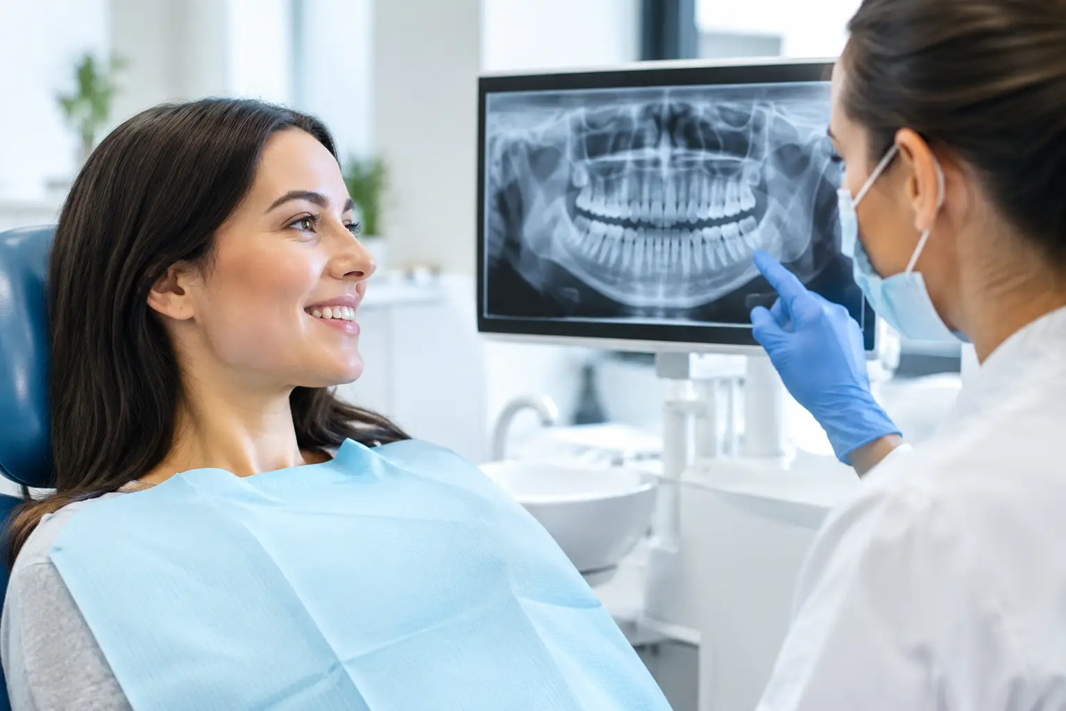
A cavity can begin between two teeth where even a careful brushing routine and a close visual exam cannot see it. Early bone changes from gum disease can be just as hidden. That is why many patients ask, are dental x rays safe before an exam: they want the reassurance of thorough care without unnecessary exposure.
For most patients, dental X-rays are considered very safe when they are taken only when clinically needed and with modern digital technology. They use a small amount of radiation, and the information they provide can help your dentist find problems early – often before they become painful, more complex, or more costly to treat.
At a patient-centered dental office, the goal is never to take images just because it is time for a routine schedule. Your dentist should consider your oral health history, current symptoms, risk for decay or gum disease, previous images, and the type of care you need. You deserve to understand why an X-ray is being recommended and to feel confident in that decision.
Are Dental X Rays Safe With Modern Technology?
Yes. Modern dental imaging is designed to keep exposure as low as reasonably achievable while still producing a clear, useful image. Digital X-rays generally require less radiation than older film-based X-rays, and they allow the dentist to view images quickly, adjust visibility when appropriate, and avoid retakes caused by film processing issues.
Radiation is measured in units called microsieverts. The amount from a single dental image is small, although the exact dose varies by the type of image, the equipment, and the settings used. A small X-ray inside the mouth has a lower dose than larger images such as panoramic X-rays, which show the full jaw. Cone beam CT scans, often used for more detailed planning, involve more radiation than standard dental X-rays but can provide valuable three-dimensional information when a regular image is not enough.
Daily life also involves natural background radiation from the ground, air, food, and other sources. Comparing every exposure can be reassuring, but the most meaningful point is this: dental X-rays should have a clear diagnostic purpose. A carefully selected image can prevent a hidden issue from progressing into an emergency.
Why Your Dentist May Recommend X-Rays
A visual exam remains essential, but it has limits. Teeth touch one another, roots sit beneath the gums, and the jawbone cannot be evaluated fully by looking in the mouth. X-rays allow your dentist to see areas that would otherwise remain unknown.
They may help identify decay between teeth, infection around a root, cysts, impacted teeth, bone loss related to periodontal disease, cracks, failing restorations, and changes beneath crowns or fillings. For children and teens, imaging can also help monitor tooth development and guide orthodontic planning. For adults considering implants, advanced imaging may be needed to assess available bone and the location of important structures before treatment.
Finding a concern early often means more conservative care. A small cavity may need a filling rather than a root canal and crown. Bone loss identified early may be addressed with focused periodontal care before teeth become loose. In this sense, avoiding a necessary X-ray can sometimes carry its own risk.
How Often Do You Need Dental X-Rays?
There is no single X-ray schedule that fits every patient. Someone with a history of frequent cavities, active gum disease, dry mouth, extensive dental work, or new symptoms may need images more often than someone with stable oral health and a low risk for decay.
Your dentist will also review any recent X-rays you have had. If you are new to the practice, bringing prior records can help prevent unnecessary duplicate images. When images are needed, the type matters too. A few targeted bitewing X-rays used to check between back teeth are different from a full-mouth series or a panoramic image used for broader evaluation.
If you are unsure, a simple question can bring clarity: “What are you looking for with this X-ray, and will it change my care?” A trustworthy dental team will welcome that conversation and explain the recommendation in plain language.
What Safety Steps Should a Dental Office Take?
Safe imaging is not only about having digital equipment. It is also about clinical judgment, proper training, and careful technique. Your dental team should use the smallest appropriate image area and the lowest exposure settings that will still provide a diagnostic result.
Positioning matters because a properly placed sensor reduces the chance that an image will need to be repeated. Protective measures may also be used based on current recommendations and your individual circumstances. The practice should maintain its equipment, follow applicable radiation safety requirements, and keep images in your record so future care can be planned thoughtfully.
For patients with dental anxiety, knowing what to expect can make the process feel easier. Most standard dental X-rays take only a few minutes. The sensor may feel a little awkward in the mouth, especially if you have a sensitive gag reflex or sore areas, but let the team know. They can slow down, adjust positioning, and guide you through it with a gentle touch.
Pregnancy and Dental X-Rays
Pregnancy is a common reason patients hesitate to have dental imaging. Routine elective X-rays may be postponed when they are not needed immediately, but pregnancy does not automatically mean that all dental X-rays must be avoided.
If you have severe pain, swelling, trauma, or signs of infection, an X-ray may be necessary to diagnose the problem and guide safe treatment. Untreated dental infection can create greater health concerns than a carefully performed, clinically necessary image. Tell your dentist if you are pregnant or think you may be pregnant so your care can be planned appropriately.
The same principle applies to children. Dental X-rays can be safe for young patients when they are justified and tailored to the child’s needs. Since children’s mouths change quickly and decay can progress faster in baby teeth, targeted imaging may sometimes be the best way to protect their developing smile.
When a 3D Dental Scan Makes Sense
Not every dental concern calls for a cone beam CT scan. Standard two-dimensional X-rays are often all that is needed for routine exams, cavity detection, and many restorative decisions. A 3D scan is typically reserved for situations where more detail will meaningfully improve planning or safety.
For example, your dentist may recommend a CBCT scan before placing an implant, evaluating an impacted wisdom tooth close to a nerve, assessing certain root canal concerns, or investigating complex jaw symptoms. Because these scans involve more radiation than routine images, the benefit should be clear and specific. The question is not whether more imaging is always better. It is whether the information will help deliver more precise, safer care.
A Reassuring Approach to Dental Imaging
It is reasonable to ask questions about radiation, especially if you or your child has had several medical or dental images recently. Honest dentistry means discussing both the benefit and the limitation of an X-ray recommendation. Sometimes the right answer is to take an image now. Other times, your dentist may decide that monitoring an area or relying on recent images is appropriate.
At Shine & Sparkle Dentistry, patients can expect personalized recommendations, modern technology, and clear explanations before care moves forward. Whether you are coming in for a routine checkup, managing tooth pain, or planning a more advanced treatment, imaging should support your comfort and your long-term oral health.
If an X-ray has been recommended, do not let uncertainty keep you from asking for an explanation. A few calm, clear answers can help you make a decision that protects both your peace of mind and your smile.